Medical Image Analysis Showcase
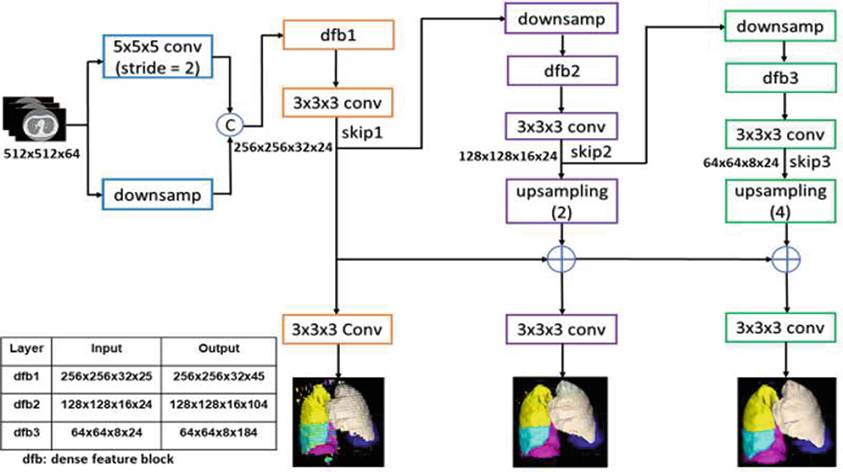 Award-winning paper on a progressive dense V-network for automatic pulmonary lobe segmentation.
Reference | PDF
 Automated pericardial fat quantification from coronary magnetic resonance angiography.
Reference | PDF
 Automated pericardium delineation and epicardial fat volume quantification from noncontrast CT.
Reference | PDF
 On the diagnosis of acute pulmonary embolism from CTPA.
Reference | PDF
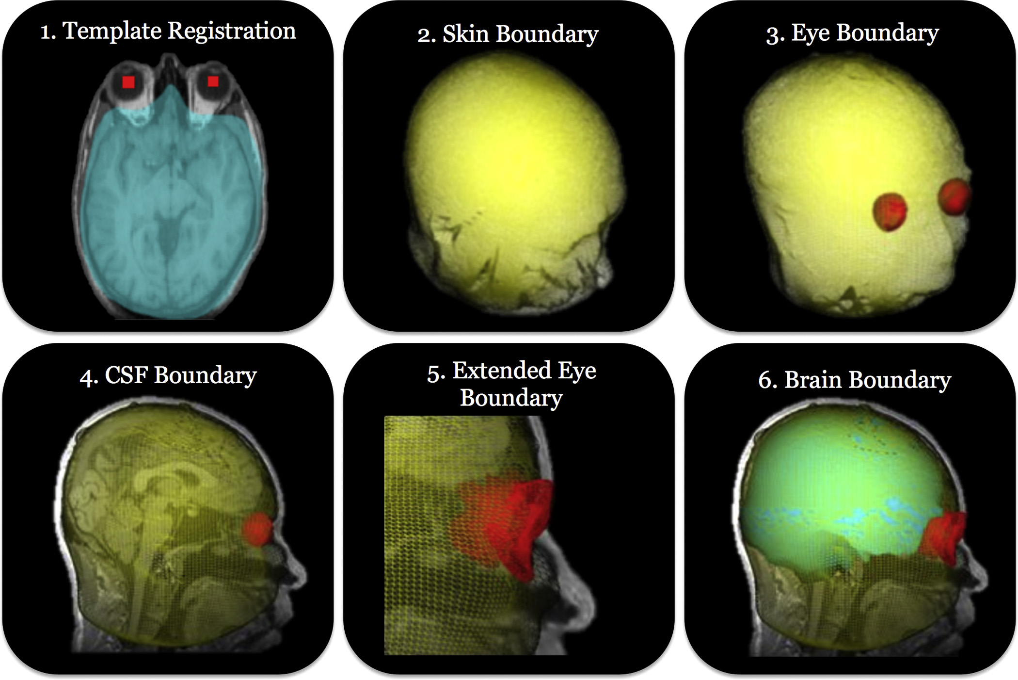 Machine learning deformable organisms applied to skull-stripping.
Reference | PDF
 MIA
journal article on "deformable organisms". The marriage of deformable
models and artificial life for automatic segmentation. Cited by the
Medical Informatics Association among The Best of Medical
Informatics, 2003.
MIA
journal article on "deformable organisms". The marriage of deformable
models and artificial life for automatic segmentation. Cited by the
Medical Informatics Association among The Best of Medical
Informatics, 2003.
Reference | PDF
Demos
A more recent book chapter on
deformable organisms
 MIA
survey on deformable models in medical image analysis. Has become a
standard reference and one of the most highly cited papers
published in this top journal.
MIA
survey on deformable models in medical image analysis. Has become a
standard reference and one of the most highly cited papers
published in this top journal.
Reference | Updated survey PDF | Original PDF | References cited in the survey, in
BibTeX format
Another
relevant survey article on nonrigid image registration
 MIA
journal article on "United Snakes", the marriage of snakes and
livewire.
MIA
journal article on "United Snakes", the marriage of snakes and
livewire.
Reference | PDF
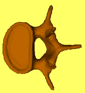
 A
multiresolution, non-intersecting deformable surface model for 3D
image segmentation/reconstruction. C&G 2001 Best Paper Award
honorable mention.
A
multiresolution, non-intersecting deformable surface model for 3D
image segmentation/reconstruction. C&G 2001 Best Paper Award
honorable mention.
Reference | PDF
 IEEE TMI journal article on
topologically adaptable surfaces (T-surfaces). A 3D generalization of
the T-snakes model below. Cited by the Medical Informatics Association
among The Best of Medical Informatics, 1999.
IEEE TMI journal article on
topologically adaptable surfaces (T-surfaces). A 3D generalization of
the T-snakes model below. Cited by the Medical Informatics Association
among The Best of Medical Informatics, 1999.
Reference
| PDF
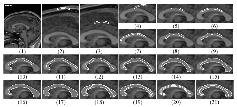
 MIA
journal article presenting topologically adaptable snakes (T-snakes)
for the analysis of medical images. A Lagrangian, active contour model
that offers a superset of the benefits of Eulerian, level-set
methods.
MIA
journal article presenting topologically adaptable snakes (T-snakes)
for the analysis of medical images. A Lagrangian, active contour model
that offers a superset of the benefits of Eulerian, level-set
methods.
Reference | PDF
ICCV'95 version of
the paper: Reference | .ps.gz
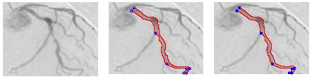
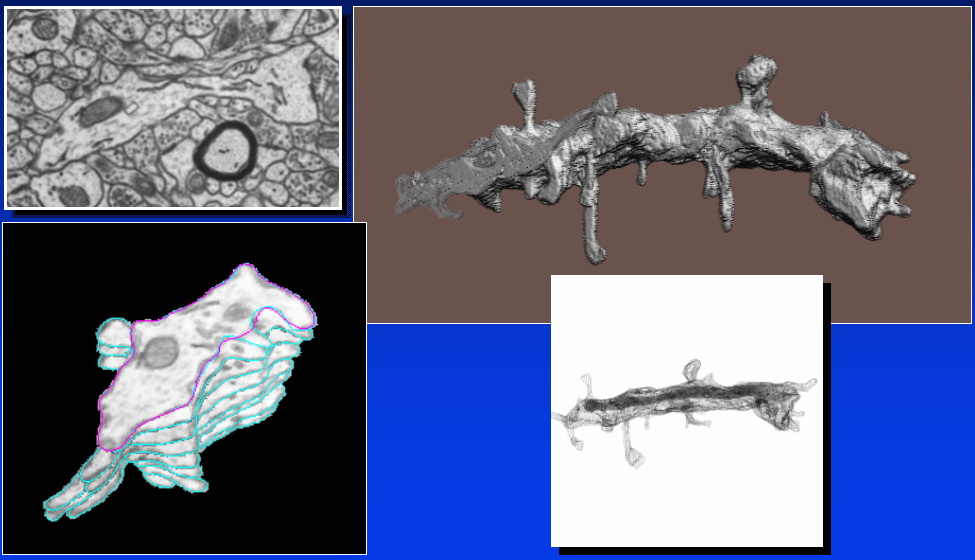 IEEE TMI
paper on fast, interactive serial segmentation of EM neuronal images
using snakes, as well as reconstruction and volume visualization of 3D
neuronal models.
IEEE TMI
paper on fast, interactive serial segmentation of EM neuronal images
using snakes, as well as reconstruction and volume visualization of 3D
neuronal models.
Reference | PDF
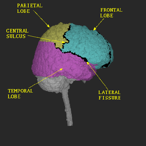
 CMIG
paper presenting a physics-based approach to anatomical surface
segmentation, reconstruction, and tracking in multidimensional medical
images using a deformable balloon model.
CMIG
paper presenting a physics-based approach to anatomical surface
segmentation, reconstruction, and tracking in multidimensional medical
images using a deformable balloon model.
Reference | PDF | .ps.gz
 My earliest research in medical image
analysis was done in 1978–1980, prior to the existence of
journals like MIA and IEEE TMI. This CGIP paper on the detection
of osteogenesis imperfecta using automated texture analysis was
published from that
work.
My earliest research in medical image
analysis was done in 1978–1980, prior to the existence of
journals like MIA and IEEE TMI. This CGIP paper on the detection
of osteogenesis imperfecta using automated texture analysis was
published from that
work.
Reference
| PDF
Demetri Terzopoulos